US-1B
;void(0);)
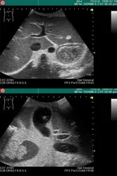
Unique high-fidelity ultrasound phantom facilitates effective training in abdominal ultrasound scanning with your own clinical devices. Simulated lesions embedded as targets provide wider educational opportunities. |
Last ned produktbeskrivelse (PDF)
Se video (WMV) 
|
Produktegenskaper
|
|
SpecificationsThe phantom includes:
Pathology includes:
Set Includes:
|
|
|
|
|
Teknisk informasjon
The classification of lung sounds is based on the criteria of the American Thoracic Society.
| 36 cases are available for training. 34 cases include 2 versions -- with and without heart-sounds | |||
|---|---|---|---|
| NORMAL | standard | FINE CRACKES | both lower area |
| mildly weak | both lower and middle area | ||
| mildly strong | whole thorax 1 | ||
| mildly rapid | whole thorax 2 | ||
| loud heart sounds | WHEEZES | upper and middle area | |
| ABNORMAL | weak: left lower area | around trachea and upper area1 | |
| weak: left whole area | around trachea and upper area2 (polyphone) | ||
| absent: left | RHONCHI | trachea and upper area | |
| weak: right lower area | trachea and upper area (polyphonic) | ||
| weak: right lower area | with an inspiratory wheeze | ||
| absent right | whole thorax | ||
| weak: whole thorax | MISCELLANEOUS CONTINUOUS SOUND |
stridor | |
| bronchial sounds | squawk | ||
| COARSE CRACKELS | right lower area | MISCELLANEOUS | pleural friction rub: left lower area |
| both lower area | pleural friction rub: right lower and middle area | ||
| right middle area | Hamman's sign | ||
| left lower area | Vocal fremitus (palpable at both sides of the chest) | ||
| both upper area | |||
| whole thorax | |||
Components & Specifications
| Component | Qty | Measurements | Packing size | Specifications |
|---|---|---|---|---|
| LSAT model unit | 1 | 32 x 35 x 62H cm | 51 x 46 x 80 cm 10 kg | Torso with rotary base 15 built-in speakers 8 ch. amplifier |
| PC | 1 | 59 x 59 x 40 cm 15 kg | Windows XP, 12ch.D/A PCI board,mouse, 112keyboard, 15"TFT monitor *Software & data installed |
|
| Amplifier | 1 | 32 x 35 x 8H cm | 46 x 46 x 15 cm 10 kg | AC 120-240V |
| Speakers | 2 | 62 x 41 x 40 cm 20 kg (incl. monitor) |
||
| T-shirt | 1 | free size |