PH-1
;void(0);)
This is a multipurpose phantom which is applicable for both plain radiography and CT scanning. The inner components consisting of mediastinum, pulmonary vasculature and an abdomen block are easily detachable, allowing insertion of mimic tumors or other lesions. The unique radiological substitute material and the elaborate three-dimensional modeling of pulmonary vessels offer the most life-like X-ray and CT images. A combination of various approaches will enrich the training opportunities.
Production Supervision:
Kiyoshi Murata, Ph.D Professor, Shiga University of Medical Science
|
Last ned produktbeskrivelse (PDF)
Se video (WMV) 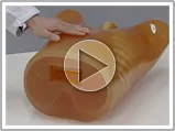
|
Produktegenskaper
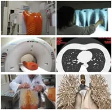
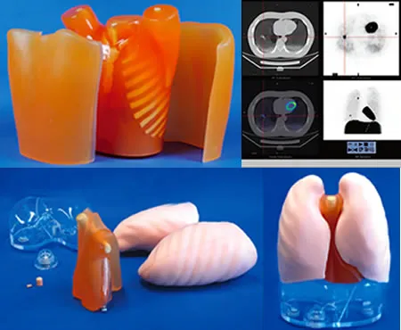
MultipurposeApplicable for both plain radiography and CT scanning. Wide variety of uses in interpretation training, anatomical education, evaluation and assessment of devices and other research.Accurate anatomy and high quality substitute materialsThe phantom is an accurate life-size anatomical model of a human torso. The thickness of the chest wall is based on measurement of clinical data. The soft tissue substitute material and synthetic bones have x-ray absorption rates very close to those of human tissues.X-rayThe phantom provides life-like radiographs very close to actual clinical images. The three-dimensional structure allows both PA and LATERAL images to be obtained. The phantom bones and vessels show life-like contrast gradations on the image along with tube voltages.Computed tomographyArms-abducted position of the torso suits the CT scanning. The pulmonary vessels are spatially traceable. Assessment of computer-aided detection systems is possible. |
|
Skills, Trainings & ApplicationsPlain radiography:
Computed tomography:
|
|
Expand N1 "LUNGMAN" with optional components
Chest Plate:
Components for Radioisotope:
|
|
SpecificationsSet includes: 1 male chest torso
Materials:
Phantom size:
Packing size:
|
|
|
|
|
Optional parts
|
Teknisk informasjon
The classification of lung sounds is based on the criteria of the American Thoracic Society.
| 36 cases are available for training. 34 cases include 2 versions -- with and without heart-sounds | |||
|---|---|---|---|
| NORMAL | standard | FINE CRACKES | both lower area |
| mildly weak | both lower and middle area | ||
| mildly strong | whole thorax 1 | ||
| mildly rapid | whole thorax 2 | ||
| loud heart sounds | WHEEZES | upper and middle area | |
| ABNORMAL | weak: left lower area | around trachea and upper area1 | |
| weak: left whole area | around trachea and upper area2 (polyphone) | ||
| absent: left | RHONCHI | trachea and upper area | |
| weak: right lower area | trachea and upper area (polyphonic) | ||
| weak: right lower area | with an inspiratory wheeze | ||
| absent right | whole thorax | ||
| weak: whole thorax | MISCELLANEOUS CONTINUOUS SOUND |
stridor | |
| bronchial sounds | squawk | ||
| COARSE CRACKELS | right lower area | MISCELLANEOUS | pleural friction rub: left lower area |
| both lower area | pleural friction rub: right lower and middle area | ||
| right middle area | Hamman's sign | ||
| left lower area | Vocal fremitus (palpable at both sides of the chest) | ||
| both upper area | |||
| whole thorax | |||
Components & Specifications
| Component | Qty | Measurements | Packing size | Specifications |
|---|---|---|---|---|
| LSAT model unit | 1 | 32 x 35 x 62H cm | 51 x 46 x 80 cm 10 kg | Torso with rotary base 15 built-in speakers 8 ch. amplifier |
| PC | 1 | 59 x 59 x 40 cm 15 kg | Windows XP, 12ch.D/A PCI board,mouse, 112keyboard, 15"TFT monitor *Software & data installed |
|
| Amplifier | 1 | 32 x 35 x 8H cm | 46 x 46 x 15 cm 10 kg | AC 120-240V |
| Speakers | 2 | 62 x 41 x 40 cm 20 kg (incl. monitor) |
||
| T-shirt | 1 | free size |