M-93UB
;void(0);)
|
Last ned produktbeskrivelse (PDF)
Se video (WMV) 
|
|
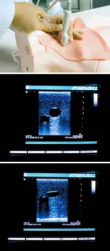
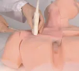
Enhanced training in ultrasound guided CVC.
Production Supervision:Akira Okada, M.D., F.A.C.S., PresidentOsaka Medical Center and Research Institute for Maternal and Child Health Masahiro Tanabe, M.D., Ph.D., Director Postgraduate and Continuing Medical Education Center Chiba University School of Medicine Kinya Sando, M.D., Ph.D., Professor Division of Human Dietetics Graduate School of Human Science Osaka Shoin Women's University Masanori Hoki, M.D., Ph.D. Head of Nutrition Management Center, Head of Community Health Service Center Chief Pediatric Surgeon Division of Pediatric Surgery Rinku General Medical Center Izumisano Municipal Hospital |
Funksjoner
Features
|
|
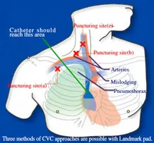
Skills & TrainingThree CVC approaches with landmark method:
Ultrasound guided CVC from internal jugular vein and axillary approach. Thorough procedure from puncture to cannulation Anatomical learning Complications indications:
|
|
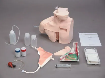
SpecificationsSet includes:
|
|
Manikin size:40 x 41 x 21H cm, 2.2 kgPacking size:52 x 46 x 39 H cm, 9 kg*Specifications are subject to change. |
|
Replacement partsReplacement parts:
|
Teknisk informasjon
The classification of lung sounds is based on the criteria of the American Thoracic Society.
| 36 cases are available for training. 34 cases include 2 versions -- with and without heart-sounds | |||
|---|---|---|---|
| NORMAL | standard | FINE CRACKES | both lower area |
| mildly weak | both lower and middle area | ||
| mildly strong | whole thorax 1 | ||
| mildly rapid | whole thorax 2 | ||
| loud heart sounds | WHEEZES | upper and middle area | |
| ABNORMAL | weak: left lower area | around trachea and upper area1 | |
| weak: left whole area | around trachea and upper area2 (polyphone) | ||
| absent: left | RHONCHI | trachea and upper area | |
| weak: right lower area | trachea and upper area (polyphonic) | ||
| weak: right lower area | with an inspiratory wheeze | ||
| absent right | whole thorax | ||
| weak: whole thorax | MISCELLANEOUS CONTINUOUS SOUND |
stridor | |
| bronchial sounds | squawk | ||
| COARSE CRACKELS | right lower area | MISCELLANEOUS | pleural friction rub: left lower area |
| both lower area | pleural friction rub: right lower and middle area | ||
| right middle area | Hamman's sign | ||
| left lower area | Vocal fremitus (palpable at both sides of the chest) | ||
| both upper area | |||
| whole thorax | |||
Components & Specifications
| Component | Qty | Measurements | Packing size | Specifications |
|---|---|---|---|---|
| LSAT model unit | 1 | 32 x 35 x 62H cm | 51 x 46 x 80 cm 10 kg | Torso with rotary base 15 built-in speakers 8 ch. amplifier |
| PC | 1 | 59 x 59 x 40 cm 15 kg | Windows XP, 12ch.D/A PCI board,mouse, 112keyboard, 15"TFT monitor *Software & data installed |
|
| Amplifier | 1 | 32 x 35 x 8H cm | 46 x 46 x 15 cm 10 kg | AC 120-240V |
| Speakers | 2 | 62 x 41 x 40 cm 20 kg (incl. monitor) |
||
| T-shirt | 1 | free size |